Label the following parts in the given diagram using arrow.
(a) Aqueous chamber (b) Cornea (c) Lens (d) Retina (e) Vitreous chamber (f) Blind spot

Representation of the following parts of eye

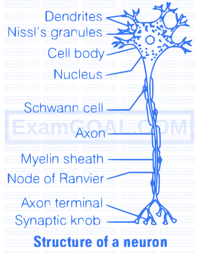
A neuron has three main parts
(i) Cell body (ii) Axon (iii) Dendrites
Any stimulus/nerve impulse passes from one neuron to another via axon. This nerve impulse is wave of bioelectric/electrochemical disturbance that passes along neuron during conduction of an excitation.
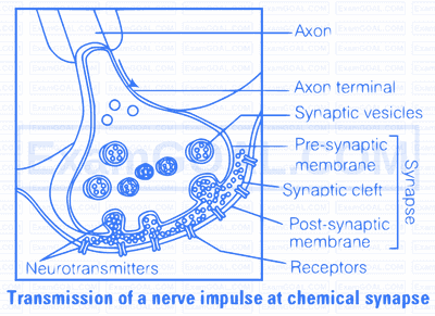
Transport and release of a neurotransmitter occurs within a synapse. At a chemical synapse, the membranes of the pre- and post-synaptic neurons are separated by a fluid-filled space called synaptic cleft. Chemicals called neurotransmitters are involved in the transmission of impulses at these synapses. The axon terminals contain vesicles filled with these neurotransmitters. When an impulse (action potential) arrives at the axon terminal, it stimulates the movement of the synaptic vesicles towards the membrane, where they fuse with the plasma membrane and release their neurotransmitters in the synaptic cleft. The released neurotransmitters bind to their specific receptors, present on the post-synaptic membrane. This binding opens ion channels allowing the entry of ions which can generate a new action potential in the post-synaptic neuron.
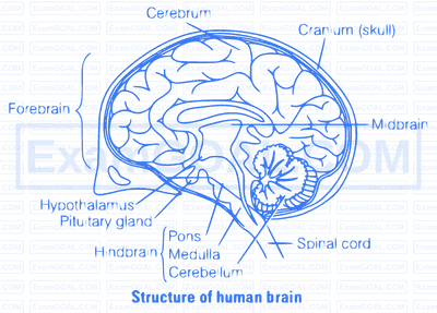
The forebrain is the largest part of the brain most of which is cerebrum. Other important structures include the thalamus, hypothalamus and the limbic system. The cerebrum is divided into two cerebral hemisphere connected here by a mass of white matter known is corpus callosum. Each hemispere is split into four lobes. The surface of each hemisphere is made up of grey matter known as the cerebral cortex that is folded to increase the surface area. Various structures of forebrain are given below
| Brain Region | Structure | Function |
|---|---|---|
| Diencephalon | Thalamus | Organising sensory information |
| Diencephalon | Hypothalamus | Endocrine system, thermoregulation |
| Diencephalon | Pituitary | Endocrine system |
| Telencephalon | Cerebral cortex | Consciousness, language, etc |
| Telencephalon | Limbic system | Memory, motivation, emotions |
| Telencephalon | Olfactory bulb / lobes | Smell |
Thalamus
The thalamus has many functions including processing and relaying sensory information selectively to various parts of the cerebral cortex, translating signals to the cerebral cortex and also regulating states of sleep and wakefulness. The thalamus plays a major role in regulating arousal levels of consciousness and levels of activity.
Hypothalamus
The function of the hypothalamus is mainly related to the overall regulation of the endocrine system and closely related to the pituitary gland.
Pituitary
The function of the pituitary is mainly related to the production of hormones as part of the endocrine system.
Cerebral Cortex
The cerebral cortex is essential for memory, attention, awareness, thought, language and consciousness. The cerebral cortex is connected to structures such as the thalamus and the basal ganglia, sending information to them along efferent connections and receiving information form them via afferent connections.
Motor Cortex
The motor cortex areas of the brain are located in both hemispheres of the cortex are related to controlling voluntary movements, especially fine movements.
Sensory Areas
The sensory areas are the areas theat receive and process information from the senses. inputs form the thalamus are called primary sensory areas, where vision, hearing and touch are processed. The two hemispheres of the cerebral cortex receive information form the opposite (contra lateral) side of the body.
The association areas of the brain function to produce a perception of the world enabling an animal to interact with their environment effectively. The frontal lobe or prefrontal association complex is involved in planning actions and movement.
Limbic System
The limbic system is principally responsible for emotions and the various types of emotion can affect the activity of the Autonomic Nervous System (ANS) facilitated by the hypothalamus.
Olfactory Bulb
The olfactory bulb is responsible for olfaction concerned with sense of small plays.
Ears are a pair of statoacoustic organ meant for both balancing and hearing. In most mammals, the external ear is a leap of tissue also called pinna. It is a part of auditory system. The human ear consists of three main parts external ear, middle, ear and internal ear.
Structure of Middle Ear
The middle ear contains three bones or ossicles-the malleus (hammer), incus (anvil) and stapes (stirr-up). These bones are attached to one another in a chain-like fashion. The malleus is attached to the tympanic membrane and the stapes is attached to the oval window (a membrane beneath the stapes) of cochlea. These three ossicles increase the efficiency of transmission of sound waves to the inner ear.
The middle ear also opens into the Eustachian tube, which connects with the pharynx and maintains the pressure between the middle ear and the outside atmosphere,
Structure of Internal Ear
The inner ear consists of a labyrinth of fluid-filled chambers within temporal bone of the skull. The labyrinth consists of two parts, i.e., the bony and membranous labyrinth. The bony labyrinth is a series of channels. Inside these channels, membranous labyrinth lies, which is surrounded by a fluid called perilymph. The membranous labyrinth is filled with a fluid called endolymph. The coiled portion of the labyrinth is called cochlea.
The cochlea has two large canals-an upper vestibular canal (scala vestibuli) and a lower tympanic canal (scala typmani)-separated by a small cochlear duct (scala media). The vestibular and tympanic canals contain perilymph and the cochlear duct is filled with endolymph.
At the base of scale vestibuli, the wall of membranous labyrinth comes in contact with the fenestra ovalis, while at the lower end of scala tympani lies the fenestra rotunda.