How does a muscle shorten during its contraction and return to its original form during relaxation?
Formation of cross-bridge between the actin and myosin filament help muscle to contract. (i) An ATP molecule joins the active site on myosin head of myosin myofilament. These heads contains an enzyme, myosin ATPase that along with $\mathrm{Ca}^{2+}$ and $\mathrm{Mg}^{2+}$ ions catalyses the breakdown of ATP. $$ \text { ATP } \xrightarrow[\mathrm{Ca}^{2+} \mathrm{Mg}^{2+}]{\text { Myosin ATPase }} \mathrm{ADP}+\mathrm{P}_j+\text { Energy } $$ (ii) The energy is transferred to myosin head which energises and straightens to join an active site on actin myofilament, forming a cross-bridge.
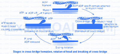
(iii) The energised cross-bridges move, causing the attached actin filaments to move towards the centre of A-band. The Z-line is also pulled inwards causing shortening of sarcomere, i.e., contraction. It is clear from the above explanation that during contraction A-bands retain the length, while I-bands get reduced.
(iv) The myosin head releases ADP and Pi, relaxes to its low energy state. The head detaches from actin myofilaments when new ATP molecule joins it and cross-bridge are broken.
(v) In repeating cycle, the free head cleaves the new ATP. The cycles of cross-bridge formation and breakage is repeated causing further sliding.

(vi) Muscle relaxation occurs after contraction when the calcium ions are pumped back to the sarcoplasmic cisternae, thus, blocking the active sites on actin myofilaments. The Z-line returns to original position, i.e., relaxation.
Calcium plays a key regulatory role in muscle contraction. These ions bind to troponin causing change in its shape and position. This in turn alters the shape and position of tropomyosin. This shift exposes the active sites on the F-actin molecules and myosin cross-bridges able to bind to these active sites.
The complete process is outlined in the figure below
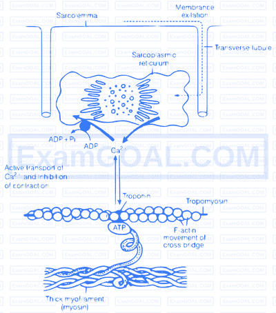
Role of calcium ion, is the contraction and relaxation process. The head of each myosin molecule contains an enzyme myosin ATPase. In the presence of myosin ATPase, $\mathrm{Ca}^{2+}$ and $\mathrm{Mg}^{2+}$ ions, ATP breaks down into ADP and inorganic phosphate as
$$\text { ATP } \xrightarrow[\mathrm{Ca}^{2+}, \mathrm{Mg}^{2+}]{\text { Myosin ATPase }} \mathrm{ADP}+\mathrm{P}_{\mathrm{i}}+\text { Energy }$$
Energy from ATP causes energised myosin cross-bridges to bind with actin.
The pectoral and pelvic girdle are responsible in providing support to the upper and lower body portions
| Pectoral Girdle | Pelvic Girdle |
|---|---|
| It occurs in the shoulder region, hence also called as shoulder girdle. | It occurs in the hip region, hence also called as hip girdle. |
| Pectoral girdles are divided into two parts, i.e., one clavicle and one scapula. | There is one pelvic girdle, which is formed by two, innominate bones. Each bone consist of three parts. i.e., ilium, ischium and pubis. |
| Image | Image |
| Clavicle and scapula helps in articulation of the upper limb with axial skeleton. | The innominate at the middle of its lateral surface has a deep, cup shaped acetabulum. where head of the femur articulates the two halves of the pelvic girdle and meet ventrally to form public symphysis. |
| It has no articulation with the vertebral column. | It has articulation with vertebral column. |
| Bones associated with pectoral girdle are light, as they are not subjected to much stress. | Bones associated with pelvic girdle are hard as they are subjected to much stress |
| There perform like holding, lifting | There function like running, standing, jumping |