The process of release of urine is called micturition and the neural mechanism. causing it is called the micturition reflex.
The urinary bladder and the internal sphincter are supplied by sympathetic and parasympathetic nervous systems of autonomic nervous system. In response, the stretch receptors on the walls of the bladder send signals to the Central Nervous System (CNS).
The CNS passes on motor messages to initiate the contraction of smooth muscles of the bladder and simultaneous relaxation of the urethral sphincter causing the release of urine.
Disorders of excretory system includes
(i) Uremia It is the malfunctioning of kidneys, which leads to accumulation of urea in blood in turn the kidney faliure. In such patients urea can be removed by haemodialysis.
(ii) Renal Failure (RF) It is caused by a decrease in glomerular filteration. In Acute Renal Failure (ARF) both kidneys abruptly stop working due to damaged renal tubules, kidney stones, antibiotics, etc. Haemodialysis and renal transplant are the only ways to auruve overcome renal failure.
(iii) Renal Calculi It is the formation of stones or insoluble mass of crystallised salts in the kidney.
(iv) Glomerulonephritis It is the inflammation of glomeruli of kidney.
In addition to the role of Proximal Convoluted Tublues (PCT) in selective reabsorption of materials from the glomerular filtrate back into the blood of peritubular capillaries via the renal interstitium, they also alter the composition of filtrate by the process of secretion.
In its distal part, epithelial cells extract certain excretory substances from the blood of peritubular capillaries and secrete these into the filtrate.
Creatinine, hippuric acid, pigments, drugs including penicillin are actively secreted into the filtrate in the proximal convolued tubule from the interstitial fluid. Hydrogen ions and ammonia are also secreted into the proximal convoluted tubules.
Urea enters the filtrate via diffusion in the thin segment of ascending limb of Henle's loop.
Maximum hydrogen secretion occurs in the proximal convoluted tubules. Removal of hydrogen ion and $\mathrm{NH}_3$ from the blood in the PCT and Distal Convoluted Tubule (DCT) helps in maintaining pH of the blood, i.e., between 6 to 8.
Tubular secretion although possess a minor role in functioning of the human kidney, but plays an essential role in animals like marine fishes and desert amphibians, because these animal do not possess well developed glomeruli hence their urine is mainly formed by the tubular secretion of urea, creatinine and mineral ions.
The glomerular filtrate in the loop of Henle gets concentrated in the descending loop and then gets diluted in the ascending limb. The thin wall of descending limb of Henle's loop is permeable to water, but not to the solutes. The isotonic tubular fluid flows down the limb.
It gradually looses its water the by exosmosis due to increasing osmolarity of medullary interstitium through which the limb extends.
Thus, the filtrate becomes hypertonic to blood plasma. The ascending limb of loop of Henle is impermeable to water, but permeable to ions like $\mathrm{K}^{+}, \mathrm{Cl}^{-}, \mathrm{Na}^{+}$and it is partially permeable to urea.
Thus, in the thick ascending limb of the loop of Henle, $\mathrm{Na}, \mathrm{K}, \mathrm{Ca}, \mathrm{Mg}$ and Cl are reabsorbed, making the filtrate hypotonic to blood plasma and diluted as compared to descending limb.
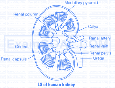
Human kidney are reddish-brown, bean-shaped structures situated between the last thoracic and third lumbar vertebra close to the dorsal inner wall of the abdominal cavity. Each kidney of an adult human measures $10-12 \mathrm{~cm}$ in length, $5-7 \mathrm{~cm}$ in width, $2-3 \mathrm{~cm}$ in thickness with an average weight of $120-170 \mathrm{gm}$.
The kidney is covered by a fibrous connective tissue, the renal capsula, which protect the kidney. Internally, it consists of outer dark cortex and an inner light medulla, both containing nephron (structural and functional units of kidney.
The median conconcave border of a kidney contains a notch called hilum. Through which ureter blood vessels and urinitus.
The renal cortex is granular in apperance and contains convoluted tubules and Malpighian corpuscles. The renal medulla contains loop of Henle, collecting ducts and tubules and ducts of Bellini.
Medulla is divided into conical masses, the medullary pyramids which further form papillae. The papillae form calyces, which join to renal pelvis leading to ureter. Between the medullary pyramids, cortex extends into medulla and forms renal columns which are called as column of Bertini.