The glomerular filtrate in the loop of Henle gets concentrated in the descending loop and then gets diluted in the ascending limb. The thin wall of descending limb of Henle's loop is permeable to water, but not to the solutes. The isotonic tubular fluid flows down the limb.
It gradually looses its water the by exosmosis due to increasing osmolarity of medullary interstitium through which the limb extends.
Thus, the filtrate becomes hypertonic to blood plasma. The ascending limb of loop of Henle is impermeable to water, but permeable to ions like $\mathrm{K}^{+}, \mathrm{Cl}^{-}, \mathrm{Na}^{+}$and it is partially permeable to urea.
Thus, in the thick ascending limb of the loop of Henle, $\mathrm{Na}, \mathrm{K}, \mathrm{Ca}, \mathrm{Mg}$ and Cl are reabsorbed, making the filtrate hypotonic to blood plasma and diluted as compared to descending limb.
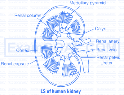
Human kidney are reddish-brown, bean-shaped structures situated between the last thoracic and third lumbar vertebra close to the dorsal inner wall of the abdominal cavity. Each kidney of an adult human measures $10-12 \mathrm{~cm}$ in length, $5-7 \mathrm{~cm}$ in width, $2-3 \mathrm{~cm}$ in thickness with an average weight of $120-170 \mathrm{gm}$.
The kidney is covered by a fibrous connective tissue, the renal capsula, which protect the kidney. Internally, it consists of outer dark cortex and an inner light medulla, both containing nephron (structural and functional units of kidney.
The median conconcave border of a kidney contains a notch called hilum. Through which ureter blood vessels and urinitus.
The renal cortex is granular in apperance and contains convoluted tubules and Malpighian corpuscles. The renal medulla contains loop of Henle, collecting ducts and tubules and ducts of Bellini.
Medulla is divided into conical masses, the medullary pyramids which further form papillae. The papillae form calyces, which join to renal pelvis leading to ureter. Between the medullary pyramids, cortex extends into medulla and forms renal columns which are called as column of Bertini.
