The cardiac cycle consist of one heart beat or one cycle of contraction and relaxation i.e., takes place in the cardiac muscles. During the heart beat there is a contraction and relaxation of atria and ventricles. The contraction phase is referred as systole while the relaxation phase is called as diastole.
The successive events of the cardiac cycle are briefly described as below
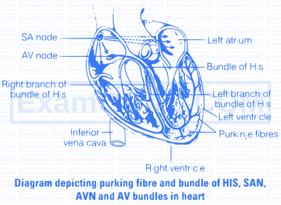
(i) Atrial Systole The atria contract due to the wave of contraction, stimulated by the SA node. The blood is forced into the ventricles as the bicuspid and tricuspid valves are open.
(ii) Beginning of Ventricular Systole The contraction of ventricles begin due to the wave of contraction stimulated by AV node. This led to the closing of bicuspid and tricuspid valve producing part of first heart sound, i.e., lub.
(iii) Complete Ventricular Systole After ventricular contraction, the blood flows into the pulmonary trunk and aorta as the semilunar valves open.

(d) Beginning of the Ventricular Diastole The ventricles relax and the semilunar valves are closed. This cause the second heart sound, i.e., dub.
(e) Complete Ventricular Diastole The opening of tricuspid and bicuspid valves due to fall in pressure of ventricles and blood flows from the atria into the ventricles. Contraction of the heart does not cause this blood to flow, backward direction, due to the fact that the pressure within the relaxed ventricles is less than that of the atria and veins.
The duration of cardiac cycle last for 0.8 sec .
In double circulation, the blood passes twice through the heart during one complete cycle. Double circulation is carried out by two ways
(i) Pulmonary circulation
(ii) Systemic circulation
Significance of Double Circulation In birds and mammals, two separate circulatory pathways are present. Oxygenated and deoxygenated blood received by the left and right atria respectively passes on to the ventricles of the same sides. The ventricles pump it out without mixing the oxygenated and deoxygenated blood in the heart.
There are more than 30 antigens on the surface of blood cells that give rise to different blood groups.
ABO Grouping ABO grouping is based on the presence or absence of two surface antigens on the RBCs namely. A and B. The plasma of different individuals contain two natural antibodies. The distribution of antigen and antibody in the four groups of blood, A , $\mathrm{AB}, \mathrm{B}$ and O are explained above in the table.
Human ABO Blood Groups and their Compatibility
| Blood Group | Genotype | Antigens on Red Blood Corpuscles | Antibodies in Blood Plasma | Donor | Recipient |
|---|---|---|---|---|---|
| A | $\mathrm{I}^A \mathrm{I}^A$ or $\mathrm{I}^A \mathrm{I}^O$ | A | b | A, AB | A, O |
| B | $\mathrm{I}^B \mathrm{I}^B$ or $\mathrm{I}^B \mathrm{I}^O$ | B | a | B, AB | B, O |
| AB | $\mathrm{I}^{\mathrm{A}} \mathrm{I}^8$ | AB | None | AB | AB, A, B, O |
| O | $\mathrm{I}^{\mathrm{O}} \mathrm{I}^O$ | None | a, b | AB, A, B, O | O |
From the above table it is evident that group ' O ' blood can be donated to persons with any other blood group and hence 'O' group individuals are called 'Universal donors'. Person with 'AB' blood can accept blood from persons with AB, as well as the other groups of blood. Therefore, such persons are called 'Universal recipients'.
Write short note on the following.
(a) Hypertension (b) Coronary Artery Disease
(a) Hypertension The high blood pressure can harm heart, brain kidneys and eyes. High blood pressure is most common disease affecting the heart and blood vessels, Blood pressure is considered normal at 120/80. When it goes beyond 140 mm Hg and 90 mm Hg it is called hypertension or high blood pressure.
Causes of Hypertension
(i) Blockage in the coronary heart vessels.
(ii) Tobacco smoking speeds up heart rate. It constrict blood vessels and raises blood pressure.
(b) Coronary Artery Disease (CAD) It is caused due to the deposition of fatty substances on the arterial wall causing atherosclerotic plaques. The lumen of artery decreases, thus obstructing the blood flow and sometimes completely blocks the artery resulting into thus, heart attack.
The diagrammatic presentation of heart with labelled SAN, AVN, AV bundles bundle of His and purkinje fibres in heart is show as follows