Write the features that distinguish between the two
(a) plasma and serum
(b) open and closed circulatory system
(c) sino-atrial node and atrio-ventricular node
(a) Difference between plasma and serum are as follows
| Blood Plasma | Blood Serum |
|---|---|
| It is the fluid without blood corpuscles. | It is liquid without dotting elements. |
| It is faint yellow in colour. | It is pale yellow. |
| It has fibrinogen and other clotting materials. | It does not have fibrinogen and other clotting materials. |
| It takes part in blood clotting. | It does not take part in blood clotting. |
(b) Difference between open and closed circulatory system are as follows
| Open Circulatory System | Closed Circulatory System |
|---|---|
| Open circulation occurs in arthropods and molluscs. | It occurs in annelids (earthworms), some molluscs and all vertebrates. |
| The blood is not completely enclosed within vessels, the heart pumps blood through arteries into large cavities or sinuses, where it mixes with the interstitial fluid and bathes the cells of the body. | In closed circulatory system, materials move between the blood and interstitial fluid through thin walls capillaries. |
| Circulation is slower in an open system, because some of the blood pooled in sinuses and, the heart is unable to build up enough pressure to make the blood flow rapidly. | Blood flows at a high pressure in a closed circulatory system. |
| Respiratory pigment, if present, is dissolved in the plasma, no red corpuscles are present. | Respiratory pigment is present which may be dissolved in the plasma but is usually contained red blood corpuscles. |
(c) Difference between sino atriol node and artrio-ventricular node are as follow
| SA Node | AV Node |
|---|---|
| It is the small mass of specialised muscle cells in the wall of right atrium near the opening of vena cava. | It is situated in the fibrous ring between the right atrium and ventricle of the heart. |
| It initiates and maintains the heart beat. | It is the pathway through, which electrical impulses can pass.s. |
Blood is a connective tissue. It has many cellular components. Thrombocytes or platelets are one of them.
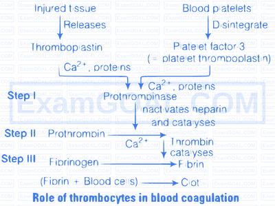
Thrombocytes or platelets are found in blood. There number in the blood is 250,000/cubic mL of blood. They are formed in bone marrow and their life span is one week. When an injury is caused in the blood vessel, bleeding starts, and the platelets are disintegrated to release the clotting factor 3 called thromboplastin. This in presence of $\mathrm{Ca}^{2+}$ ions activate prothrombokinase. A series of reactions ultimately occurs which causes blood to clot and plugg the injured blood vessel thus preventing further loss of blood.

Answer the following
(a) name the major site where RBCs are formed.
(b) which part of heart is responsible for initating and maintaining its rhythmic activity?
(c) what is specific in the heart of crocodiles among reptilians?
(a) Bone marrow
(b) SA Node (Sino Atrial Node)
(c) Reptile have 3 chambered heart with an exception of crocodile which possess 4 chambered heart, due to the partial division of ventricle through a septum.
Rh antigen is observed on the surface of RBCs of majority (nearly 80\%) of humans. Such individuals are called Rh positive $\left(\mathrm{Rh}^{+}\right)$and those individuals where this antigen is absent are called Rh negative $\left(\mathrm{Rh}^{-}\right)$.
Both $\mathrm{Rh}^{+}$and $\mathrm{Rh}^{-}$individuals are phenotypically normal. The problem in them arises during blood transfusion and pregnancy.
(i) Incompatibility During Blood Transfusion The first blood transfusion of $\mathrm{Rh}^{+}$blood to the person with $\mathrm{Rh}^{-}$blood causes no harm because the $\mathrm{Rh}^{-}$person develops anti Rh factors or antibodies in his/her blood.
In second blood transfusion of $\mathrm{Rh}^{+}$blood to the $\mathrm{Rh}^{-}$person, the already formed anti Rh factors attack and destroy the red blood corpuscles of the donor.
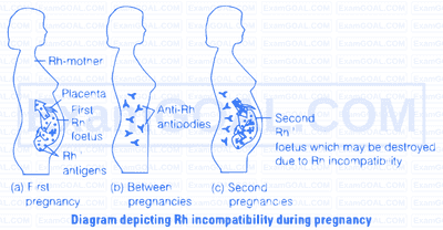
(ii) Incompatibility During Pregnancy If father's blood is $\mathrm{Rh}^{+}$, mother blood is $\mathrm{Rh}^{-}$and the foetus blood is $\mathrm{Rh}^{+}$. it will lead to a serious problem. Rh antigens of the foetus do not get exposed to the $\mathrm{Rh}^{-}$ve blood of the mother in the first pregnancy as the two bloods are well separated by the placenta.
But in the subsequent $\mathrm{Rh}^{+}$foetus, the anti Rhfactors (antibodies) of the mother destroy the foetal red blood corpuscles due to mixing of blood. This result in the Haemolytic Disease of the New Born (HDN), called as erythroblastosis foetalis. In some cases new born may survive but will be anaemic and may also suffer with jaundice.

This condition can be avoided by administering anit-Rh antibodies to the mother immediately after the delivery of the first child.
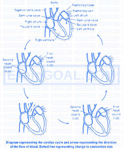
The cardiac cycle consist of one heart beat or one cycle of contraction and relaxation i.e., takes place in the cardiac muscles. During the heart beat there is a contraction and relaxation of atria and ventricles. The contraction phase is referred as systole while the relaxation phase is called as diastole.
The successive events of the cardiac cycle are briefly described as below
(i) Atrial Systole The atria contract due to the wave of contraction, stimulated by the SA node. The blood is forced into the ventricles as the bicuspid and tricuspid valves are open.
(ii) Beginning of Ventricular Systole The contraction of ventricles begin due to the wave of contraction stimulated by AV node. This led to the closing of bicuspid and tricuspid valve producing part of first heart sound, i.e., lub.
(iii) Complete Ventricular Systole After ventricular contraction, the blood flows into the pulmonary trunk and aorta as the semilunar valves open.

(d) Beginning of the Ventricular Diastole The ventricles relax and the semilunar valves are closed. This cause the second heart sound, i.e., dub.
(e) Complete Ventricular Diastole The opening of tricuspid and bicuspid valves due to fall in pressure of ventricles and blood flows from the atria into the ventricles. Contraction of the heart does not cause this blood to flow, backward direction, due to the fact that the pressure within the relaxed ventricles is less than that of the atria and veins.
The duration of cardiac cycle last for 0.8 sec .
In double circulation, the blood passes twice through the heart during one complete cycle. Double circulation is carried out by two ways
(i) Pulmonary circulation
(ii) Systemic circulation
Significance of Double Circulation In birds and mammals, two separate circulatory pathways are present. Oxygenated and deoxygenated blood received by the left and right atria respectively passes on to the ventricles of the same sides. The ventricles pump it out without mixing the oxygenated and deoxygenated blood in the heart.